
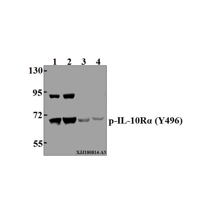
IL-10Rα (phospho-Y496) polyclonal, anti-human, mouse, rat
€328.00
In stock
SKU
BS4592
Background:
The IL-10 receptor, IL-10R, is a member of the class II subgroup of the cytokine receptor family and exhibits structural similarity to the interferon receptor. IL-10R is expressed in B cells and T helper cells, as well as in LPS-induced mouse fibroblasts. Overall, mouse IL-10R and human IL-10R share 60% sequence identity at the protein level. Stimulation with IL-10 leads to phosphorylation of JAK1 and Tyk 2 tyrosine kinases. The activated kinases phosphorylate the two tyrosine residues (Tyr 446 and Tyr 496) in the cytoplasmic domain of IL-10Rα. The phosphorylation of these two residues are required for proper function of IL-10R and activation of IL-10E1 signaling. IL-10Rβ is ubiquitously expressed and, in addition to forming the IL-10 heterodimeric receptor, it forms a heterodimeric receptor with an IL-22R subunit and an IL-28R subunit. IL-10R is constitutively expressed on human natural killer (NK) cells and the direct binding of IL-10 potentiates cytokine production by human NK cells.
Alternative Name:
IL-10R-A, IL-10R1, CDw210a antigen, IL10RA, IL10R
Application Dilution: WB: 1:500~1:1000
Specificity: p-IL-10Rα (Y496) pAb detects endogenous levels of IL-10Rα protein only when phosphorylated at Tyr496.
Immunogen:
Synthetic phosphopeptide derived from human IL-10Rα around the phosphorylation site of Tyrosine 496
MW: ~ 70 kDa
Swis Prot.: Q13651
Purification & Purity:
The antibody was affinity-purified from rabbit antiserum by affinity-chromatography using epitope-specific immunogen and the purity is > 95% (by SDS-PAGE).
Format:
1 mg/ml in Phosphate buffered saline (PBS) with 0.05% sodium azide, approx. pH 7.3.
Storage:
Store at 4°C short term. Aliquot and store at -20°C long term. Avoid freeze-thaw cycles.
For research use only, not for use in diagnostic procedure.
The IL-10 receptor, IL-10R, is a member of the class II subgroup of the cytokine receptor family and exhibits structural similarity to the interferon receptor. IL-10R is expressed in B cells and T helper cells, as well as in LPS-induced mouse fibroblasts. Overall, mouse IL-10R and human IL-10R share 60% sequence identity at the protein level. Stimulation with IL-10 leads to phosphorylation of JAK1 and Tyk 2 tyrosine kinases. The activated kinases phosphorylate the two tyrosine residues (Tyr 446 and Tyr 496) in the cytoplasmic domain of IL-10Rα. The phosphorylation of these two residues are required for proper function of IL-10R and activation of IL-10E1 signaling. IL-10Rβ is ubiquitously expressed and, in addition to forming the IL-10 heterodimeric receptor, it forms a heterodimeric receptor with an IL-22R subunit and an IL-28R subunit. IL-10R is constitutively expressed on human natural killer (NK) cells and the direct binding of IL-10 potentiates cytokine production by human NK cells.
Alternative Name:
IL-10R-A, IL-10R1, CDw210a antigen, IL10RA, IL10R
Application Dilution: WB: 1:500~1:1000
Specificity: p-IL-10Rα (Y496) pAb detects endogenous levels of IL-10Rα protein only when phosphorylated at Tyr496.
Immunogen:
Synthetic phosphopeptide derived from human IL-10Rα around the phosphorylation site of Tyrosine 496
MW: ~ 70 kDa
Swis Prot.: Q13651
Purification & Purity:
The antibody was affinity-purified from rabbit antiserum by affinity-chromatography using epitope-specific immunogen and the purity is > 95% (by SDS-PAGE).
Format:
1 mg/ml in Phosphate buffered saline (PBS) with 0.05% sodium azide, approx. pH 7.3.
Storage:
Store at 4°C short term. Aliquot and store at -20°C long term. Avoid freeze-thaw cycles.
For research use only, not for use in diagnostic procedure.
| Is Featured? | No |
|---|
Write Your Own Review