
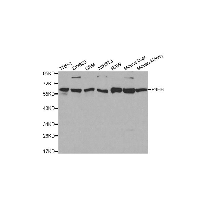
P4HB polyclonal, anti-human, mouse
€388.00
In stock
SKU
BS6048
Background:
Oxidoreductase-protein disulfide isomerase (PDI) is a homodimer consisting of subunits that catalyzes thiol-disulfide exchange, mediates folding of newly synthesized proteins and functions as a molecular chaperone. PDI localizes to the lumen of the endoplasmic reticulum (ER), where in conjunction with folding-helper proteins, such as immunoglobulin heavy chain binding protein (BiP), mediates tertiary and quaternary protein-processing. Cell surface PDI induces sulfhydryl-mediated conformational changes in integrin-mediated adhesion receptor-ligand interactions, thereby regulating integrin responses and cell adhesion. Additionally, PDI functions as a subunit of two more complex enzyme systems: the prolyl-4-hydroxylase and the triacylglycerol transfer proteins.
Alternative Name:
Cellular thyroid hormone binding protein, Cellular thyroid hormone-binding protein, Collagen prolyl 4 hydroxylase beta, Disulphide Isomerase, DSI, EC 5.3.4.1, Endoplasmic reticulum resident protein 59, ER protein 59, ERBA2L, ERp59, GIT, Gltathione insulin transhydrogenase, Glutathione insulin transhydrogenase, P4HB, P4Hbeta, p55, PDI, PDIA1, PDIA1, PDIR, PHDB, PO4DB, PO4HB, Procollagen proline 2 oxoglutarate 4 dioxygenase (proline 4 hydroxylase) beta polypeptide (pro
Application Dilution: WB: 1:500 - 1:2000, IF: 1:20 - 1:100, IP: 1:50 - 1:100
Specificity: P4HB polyclonal antibody detects endogenous levels of P4HB protein.
Immunogen:
Recombinant full length Human P4HB.
MW: ~ 57 kDa
Swis Prot.: P07237
Purification & Purity:
The antibody was affinity-purified from rabbit antiserum by affinity-chromatography using epitope-specific immunogen and the purity is > 95% (by SDS-PAGE).
Format:
1mg/ml in PBS with 0.1% Sodium Azide, 50% Glycerol.
Storage:
Store at 4°C short term. Aliquot and store at -20°C long term. Avoid freeze-thaw cycles.
For research use only, not for use in diagnostic procedure.
Oxidoreductase-protein disulfide isomerase (PDI) is a homodimer consisting of subunits that catalyzes thiol-disulfide exchange, mediates folding of newly synthesized proteins and functions as a molecular chaperone. PDI localizes to the lumen of the endoplasmic reticulum (ER), where in conjunction with folding-helper proteins, such as immunoglobulin heavy chain binding protein (BiP), mediates tertiary and quaternary protein-processing. Cell surface PDI induces sulfhydryl-mediated conformational changes in integrin-mediated adhesion receptor-ligand interactions, thereby regulating integrin responses and cell adhesion. Additionally, PDI functions as a subunit of two more complex enzyme systems: the prolyl-4-hydroxylase and the triacylglycerol transfer proteins.
Alternative Name:
Cellular thyroid hormone binding protein, Cellular thyroid hormone-binding protein, Collagen prolyl 4 hydroxylase beta, Disulphide Isomerase, DSI, EC 5.3.4.1, Endoplasmic reticulum resident protein 59, ER protein 59, ERBA2L, ERp59, GIT, Gltathione insulin transhydrogenase, Glutathione insulin transhydrogenase, P4HB, P4Hbeta, p55, PDI, PDIA1, PDIA1, PDIR, PHDB, PO4DB, PO4HB, Procollagen proline 2 oxoglutarate 4 dioxygenase (proline 4 hydroxylase) beta polypeptide (pro
Application Dilution: WB: 1:500 - 1:2000, IF: 1:20 - 1:100, IP: 1:50 - 1:100
Specificity: P4HB polyclonal antibody detects endogenous levels of P4HB protein.
Immunogen:
Recombinant full length Human P4HB.
MW: ~ 57 kDa
Swis Prot.: P07237
Purification & Purity:
The antibody was affinity-purified from rabbit antiserum by affinity-chromatography using epitope-specific immunogen and the purity is > 95% (by SDS-PAGE).
Format:
1mg/ml in PBS with 0.1% Sodium Azide, 50% Glycerol.
Storage:
Store at 4°C short term. Aliquot and store at -20°C long term. Avoid freeze-thaw cycles.
For research use only, not for use in diagnostic procedure.
| Is Featured? | No |
|---|
Write Your Own Review